CT Scan for Kidney Stones: Diagnosis & Imaging
A CT scan for kidney stones employs a rapid series of X-rays to create detailed cross-sectional views of the abdomen. Clinicians often refer to it as a CAT scan in emergency rooms and outpatient clinics. This is due to how the slices are reconstructed into clear images.
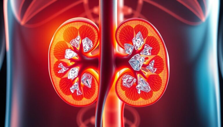
CT imaging stands as the gold standard for detecting kidney stones. It accurately measures their size and maps their location from the kidney to the bladder base. A standard kidney stone CT scan covers the upper poles of the kidneys to the bladder. This allows radiologists to identify stones, signs of obstruction, or other causes of pain like appendicitis or diverticulitis.
Speed and availability make CT the primary test for many acute flank pain cases. The drawback is radiation exposure. Yet, modern low-dose and ultra-low-dose protocols significantly reduce this risk while maintaining high accuracy. Physicians must balance diagnostic certainty with dose reduction when choosing a protocol.
What Is A CT Scan And Why It’s Used For Kidney Stones
A CT scan creates detailed images of the body by layering X-ray slices. It’s essential when someone experiences sudden flank pain or blood in their urine. This imaging technique reveals calcifications, soft tissue, and surrounding anatomy, surpassing other diagnostic methods.
CT scanners use an X-ray source and detectors that rotate around the patient. A computer then combines these images into detailed slices and reformatted views. Patients are positioned on a table that moves through the scanner, with exams typically lasting 10–20 minutes.
Radiologists use CT scans to measure stone size, location, and density in Hounsfield units. These measurements help predict if a stone can be broken with shock wave lithotripsy or if it needs ureteroscopy. Dual-energy and high-speed CT scans also provide insights into stone composition, like distinguishing between uric acid and calcium stones.
Research indicates that a renal stone CT scan can detect more than 95% of stones. It also rules out stones in over 98% of cases, as shown in many pooled reviews. This high accuracy is why emergency departments and urology services often choose CT scans for kidney stones when quick answers are critical.
Compared to ultrasound and plain X-ray, CT scans offer faster results, wider availability, and higher sensitivity. They can spot even small stones, identify obstructions, and reveal other conditions like appendicitis or diverticulitis. Low-dose protocols are used to minimize radiation while maintaining diagnostic quality for most stone evaluations.
ct scan for kidney stones
The decision to order a kidney stone ct scan is based on symptoms and clinical judgment. Sharp pain, blood in the urine, or findings suggesting renal colic often lead to CT scans. This method is favored in emergency departments for its ability to quickly distinguish stones from other causes of pain.
When Doctors Order A Kidney Stone CT Scan
Doctors opt for a ct scan when initial evaluations are unclear or symptoms are severe. Triggers include sudden flank pain, visible blood in the urine, fever with suspected infection, or when home treatments fail. The scan confirms stone presence, measures size, and guides further care.
Clinical Situations Where CT Is Preferred
Acute flank pain with suspected obstruction is the primary reason for CT scans. This modality detects small stones and identifies complications like hydronephrosis or perinephric stranding. It also helps rule out appendicitis or diverticulitis by evaluating the broader abdomen.
Recurrent episodes with unclear anatomy or previous failed conservative therapy also prompt CT scans. Surgeons and urologists rely on these scans to plan interventions like ureteroscopy or shock wave lithotripsy. They provide accurate size and location data.
Alternatives For Specific Patients
Pregnant patients typically begin with ultrasound to avoid fetal radiation. If ultrasound is inconclusive and the clinical need is high, a low-dose non-contrast CT may be considered with obstetric consultation. National health services guidelines advocate for this stepwise approach.
Children and young adults often start with ultrasound due to radiation concerns. If ultrasound is uncertain, a low-dose ct scan for kidney stones procedure can be used. Pediatric radiology teams aim to balance diagnostic yield with dose reduction to protect long-term health.
Types Of CT Scans Used For Kidney Stones
Radiologists select various CT techniques based on clinical needs. Each method balances image quality, speed, and radiation exposure. The chosen technique impacts diagnosis, follow-up, and treatment planning for kidney stone patients.
Non-Contrast CT Versus Contrast-Enhanced CT
Non-contrast CT, known as the stone protocol, is the primary test for detecting stones. It highlights stones on unenhanced images, making it quick and reliable for acute flank pain.
Contrast-enhanced CT, including CT urography for kidney stones, adds urinary tract opacification. It also reveals soft-tissue abnormalities. This is beneficial when anatomy, tumors, or unexplained blood in the urine are concerns.
Low-Dose CT And Ultra-Low-Dose CT: Definitions And Uses
Low-dose CT reduces radiation while detecting most clinically relevant stones. The effective dose is typically near 3 mSv, depending on the scanner and settings.
Ultra-low-dose CT further reduces exposure. Its sensitivity varies but is useful for repeat imaging or in younger patients at lower risk. Facilities consider the balance between dose and the risk of missing small stones.
High-Speed And Dual-Energy CT For Uric Acid Stone Detection
High-speed scanners reduce acquisition time and motion blur. They are beneficial in emergency settings and for patients who have trouble staying motionless.
Dual-energy CT distinguishes materials by energy response. It helps radiologists differentiate uric acid stones from calcium stones. This guides decisions on medical dissolution versus surgical removal.
| CT Type | Primary Strength | Typical Dose Range | Best Clinical Use |
|---|---|---|---|
| Non-Contrast CT (Stone Protocol) | High sensitivity for most calculi | Standard ~3–8 mSv (varies) | Initial diagnosis of acute flank pain and obstruction |
| Contrast-Enhanced CT / CT Urography Kidney Stones | Urinary tract opacification; soft-tissue detail | Higher than non-contrast; depends on phases | Hematuria workup, complex anatomy, tumor exclusion |
| Low-Dose CT | Lower radiation with preserved accuracy | Approximately ~1–3 mSv | Follow-up imaging and repeat studies |
| Ultra-Low-Dose CT | Minimized exposure for select patients | Below ~1 mSv in some protocols | Younger patients and frequent monitoring |
| High-Speed CT | Short acquisition time; less motion artifact | Comparable to corresponding protocol | Emergency departments and restless patients |
| Dual-Energy CT | Material characterization (e.g., uric acid) | Varies; can match standard dose | Planning medical dissolution or tailored therapy |
Radiology teams aim to optimize image quality for kidney stone CT imaging while minimizing exposure for patients needing repeat scans. The right CT type enhances diagnostic confidence and guides appropriate care.
CT Urography Kidney Stones And Specialized Protocols
CT urography offers a more detailed view than a simple non-contrast CT scan for kidney stones. It uses IV contrast and timed phases to show the collecting system, ureters, and bladder in high detail. This method is chosen when a plain CT scan leaves questions about anatomy, filling defects, or bleeding causes.
What CT Urography Adds To Standard Stone Imaging
CT urography maps contrast flow through the urinary tract. It helps spot urothelial lesions, strictures, and subtle extrinsic compression that a non-contrast CT scan might miss. Multi-phase images capture cortical enhancement, excretory patterns, and delayed filling, improving mass versus stone characterization.
When CT Urography Is Recommended
CT urography is considered in specific situations. Unexplained hematuria or suspicion of a urinary tract tumor are common triggers. Complex or congenital urinary tract anatomy, recurrent stones with suspected structural causes, and unclear obstruction etiology also warrant the study.
Before ordering CT urography, teams review renal function and allergy history. Contrast adds diagnostic value but raises concerns for patients with reduced eGFR or prior contrast reactions. Hydration protocols, alternative imaging, or adjusted timing help manage those risks.
- Unexplained hematuria: evaluate for urothelial tumor or bleeding source.
- Complex anatomy: define duplicated systems, strictures, or surgical planning.
- Recurrent stones with structural causes: check for obstruction or focal narrowing.
- Indeterminate findings on a ct scan for kidney stones: clarify filling defects.
CT urography sits alongside low-dose and dual-energy protocols in the imaging toolkit. Each choice balances diagnostic yield against radiation and contrast exposure. When precise anatomy or lesion characterization matters, CT urography for kidney stones delivers the extra detail clinicians need for confident decisions.
How To Prepare For A Kidney Stone CT Scan
Getting ready for a renal stone ct scan involves simple steps to reduce stress and ensure a smooth process. A quick checklist covers what to wear, fasting requirements, medication notes, and a brief overview of your kidney health. Clear instructions help technologists obtain the necessary images efficiently, speeding up the ct scan for kidney stones procedure.
Pre-Scan Instructions: Clothing, Jewelry, Fasting, Medications
Patients typically wear a hospital gown and remove jewelry, watches, and metal fasteners. Metal can interfere with the scan, causing artifacts that obscure the images. It’s important to bring a list of current medications and any previous imaging on a USB or phone if possible.
For non-contrast studies, fasting is usually not required. When contrast or CT urography is involved, facilities may ask for a four to six hour food avoidance. Always follow the specific instructions provided by the imaging center.
Contrast Preparation And Allergy Screening When Applicable
If IV contrast is planned, staff will inquire about any previous allergic reactions, asthma, or contrast issues. Patients with a history of contrast allergy may receive pre-medication, such as steroids or antihistamines, under a documented protocol.
Informed consent and an explanation of risks are standard. Discuss any recent reactions to iodine-based agents and medications that raise concern.
Special Considerations For Renal Impairment And Diabetes
Assessing kidney function is critical before administering IV contrast. Recent serum creatinine or eGFR results guide the team. Patients with significant renal impairment might be directed to alternative imaging or receive hydration protocols.
Diabetic patients on metformin should adhere to facility rules, which often involve holding metformin until kidney function is checked. Clear communication with the ordering clinician is essential to avoid delays in imaging.
Use the short checklist below to prepare for your appointment and reduce surprises on the day of the renal stone ct scan.
| Item | Why It Matters | Action |
|---|---|---|
| Clothing & Metal | Metal can blur images and mimic stones | Wear loose clothes; remove jewelry and belts |
| Medication List | Important for contrast safety and emergencies | Bring a current list and any allergy history |
| Fasting Rules | Affects contrast administration and nausea risk | Follow fasting instructions if contrast is planned |
| Kidney Function Tests | Determines contrast safety for the ct scan for kidney stones procedure | Provide recent creatinine/eGFR or get labs before scan |
| Contrast Allergy History | Identifies need for pre-medication or alternatives | Report prior reactions, asthma, or iodine issues |
| Diabetes & Metformin | Metformin with contrast may require temporary hold | Confirm facility protocol and pause medication if advised |
What To Expect During The CT Scan For Kidney Stones Procedure
A kidney stone CT scan is quick and often less time-consuming than patients expect. Staff will verify your registration and medical history. They will also inquire about allergies and kidney function. A brief screening determines whether a non-contrast or contrast-enhanced CT scan is necessary.
Step-By-Step: Arrival, Changing, Positioning, Imaging Time
Upon arrival, a few minutes are spent on registration and screening. You will then change into a gown and remove any jewelry. A technologist will assist in positioning you on the CT table, usually lying on your back. Thin slices are taken from the kidneys to the bladder.
Non-contrast kidney stone CT scans typically last 10–20 minutes from start to finish. Contrast studies require additional time for IV placement, injection, and any delayed phases. The technologist will inform you of the expected duration before starting the scan.
Breath-Holding, Communication With The Technologist, Noise And Comfort
Short breath-holds are sometimes requested to minimize motion blur. The scanner emits rhythmic whirring and clicking sounds during image capture. Technologists maintain constant communication via intercom and can pause the scan if needed.
Comfort measures include the use of pillows or straps to help remain steady. If claustrophobia is a concern, discussing mild anxiolytics with your clinician before arrival can help ease your experience.
Use Of IV Or Oral Contrast When Needed And Post-Scan Observation
If contrast is needed, an IV is placed, and scans are timed for specific phases. Oral contrast is rare but used when bowel detail is necessary for diagnosis. Post-contrast observation is brief for most patients.
Patients with known contrast allergies or reduced kidney function may require extended monitoring, typically 15–30 minutes. For non-contrast exams, discharge is usually immediate with routine aftercare instructions from the technologist or nurse.
| Step | Typical Time | What To Expect |
|---|---|---|
| Registration & Screening | 5–10 minutes | Medical questions, allergy and medication checks |
| Changing & Positioning | 5 minutes | Gown, remove metal, lie supine on CT table |
| Non-Contrast Imaging | 10–20 minutes total | Thin-slice acquisition from kidneys to bladder |
| Contrast-Enhanced Study | 30–60 minutes | IV placement, timed phases, possible delayed images |
| Post-Scan Observation | 0–30 minutes | Brief monitoring after contrast; non-contrast patients often leave immediately |
How CT Scan For Kidney Stones Results Are Interpreted
The radiology report transforms images into actionable insights. After a CT scan for kidney stones, radiologists detail measurements, density, and critical signs for urologists. Clear language in the report is key for quick decisions on pain management, stenting, or definitive treatments.
Reading Size, Location, Density (HU) And Clinical Implications
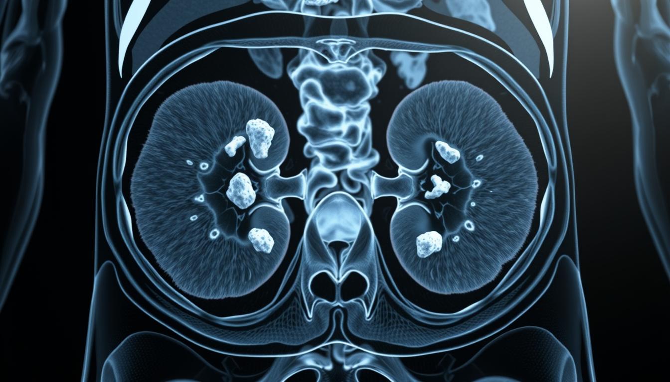
Reports outline stone diameter in millimeters and pinpoint its exact location, like renal calyx, pelvis, or ureter segments. Stone size is a critical factor; stones under 5 mm might pass naturally, whereas larger ones may require intervention.
Hounsfield units (HU) are numeric values on kidney stone CT imaging. Lower HU values suggest uric acid stones, which may dissolve with medical treatment. Higher HU values indicate calcium-rich stones, often resistant to shockwave lithotripsy.
Reporting Obstruction, Hydronephrosis, And Alternative Diagnoses
Reports discuss hydronephrosis, perinephric stranding, and ureteral impaction. Documenting obstruction severity is vital in emergency settings. Normal collecting system appearance indicates no obstruction.
CT scans can also uncover causes like appendicitis, diverticulitis, or abdominal masses. A detailed report distinguishes stone findings from other diagnoses, aiding the referring clinician in planning the next steps.
Timing Of Results And Communicating Findings To The Referring Clinician
In emergency departments, preliminary reads are often shared urgently via phone or secure message. Finalized results, including measurements, HU values, and impressions, follow within hours.
Quick and precise communication between radiologists and the treating team ensures that CT data lead to timely care. Clinicians rely on these reports to decide between conservative management or urology intervention.
Accuracy And Limitations Of Kidney Stone CT Imaging
CT imaging for kidney stones provides unparalleled clarity. It accurately maps stone size, position, and density quickly. This precision is critical for urgent medical decisions. Yet, each test comes with its own set of trade-offs.
Sensitivity And Specificity Data For Standard And Low-Dose CT
Standard non-contrast CT scans boast sensitivity near 95% and specificity around 98% in many studies. Low-dose CT scans maintain high accuracy, with sensitivity between 90% and 98%.
Ultra-low-dose techniques show sensitivity ranging from 72% to 99%. This range is influenced by patient size and scanner model. It’s important when deciding if a kidney stone ct scan will meet the diagnostic needs of surgeons or urologists.
Limitations: Very Small Stones, Radiation Trade-Offs, Cost Considerations
Stones smaller than 3 mm can be missed, often due to bowel gas or dense stool. Stones made of low-attenuation material may also be harder to spot on standard scans.
Radiation is a significant limitation. CT scans expose patients to more radiation than ultrasound or plain X-ray. This is why repeated scans increase cumulative exposure, leading to the preference for ultrasound in pregnant patients and children.
Cost and access are also factors. Not all facilities offer advanced options like dual-energy or ultra-low-dose scans. A strategy for kidney stone imaging must consider price, availability, and the need for diagnostic accuracy.
Clinicians weigh the need for diagnostic certainty against the risks of radiation and cost. In cases where anatomy or alternative diagnoses are uncertain, a renal stone ct scan is chosen. For patients at lower risk, ultrasound is the preferred initial choice.
Risks And Radiation Concerns With Renal Stone CT Scan
CT scans for kidney stones provide quick, clear results. Yet, they come with risks from ionizing radiation and contrast agents. This guide explains typical doses, long-term effects, and how to minimize harm.
The term ct scan for kidney stones radiation refers to the X-ray energy used during a scan. Older standard abdominal CT exams measured about 10 mSv in many published reports. Modern scanners and protocol tweaks often bring standard studies down. Low-dose CT protocols generally range near 3 mSv or less, depending on the machine and settings. Ultra-low-dose approaches lower exposure further but may trade off some sensitivity for very small stones.
Concerns about ct scan for kidney stones risks extend to cumulative exposure. Repeated CTs over years have been linked in some reviews to a small increased lifetime risk for certain cancers, such as thyroid malignancies or leukemia, when scans are frequent. Clinicians balance that risk against the immediate need to diagnose a suspected obstruction or severe pain.
To limit risk, teams use several strategies. They select low-dose protocols when appropriate, reserve CT for clear indications, and substitute ultrasound for follow-up when feasible. Tailoring the scan field to the area of interest and using iterative reconstruction or dual-energy scanning help lower dose without losing diagnostic value.
Contrast agents add another set of considerations. Intravenous iodinated contrast can trigger allergic reactions in a minority of patients. It may also affect renal function in people with reduced eGFR. For many stone studies, a non-contrast CT avoids those issues altogether. When contrast is needed, staff check prior reactions, measure kidney function, advise hydration, and follow metformin guidance to reduce adverse outcomes.
Below is a compact comparison to clarify typical exposures, sensitivity tradeoffs, and the main mitigation steps.
| Aspect | Standard CT | Low-Dose CT | Ultra-Low-Dose CT |
|---|---|---|---|
| Typical Radiation | Approximately 8–12 mSv on older protocols | Roughly 1.5–3 mSv with modern settings | Often below 1–1.5 mSv depending on equipment |
| Sensitivity For Stones | Very high for stones ≥2 mm | High for clinically relevant stones; slight drop for very small stones | Variable; best for larger stones, reduced accuracy for |
| When Preferred | Acute flank pain with suspected obstruction | Routine diagnosis when minimizing dose is important | Follow-up or screening in low-risk contexts |
| Contrast Use | Often non-contrast for stones; contrast used for complex cases | Usually non-contrast; contrast reserved for specific indications | Typically non-contrast to avoid added renal risk |
| Risk Mitigation | Limit repeat scans; use tailored protocols | Employ iterative reconstruction and dose modulation | Use only when clinically justified; consider ultrasound alternative |
CT Scan For Kidney Stones Cost And Insurance Considerations
Choosing imaging options often depends on cost and insurance coverage. The cost of a CT scan for kidney stones varies widely across the United States. Different settings, such as emergency rooms, hospital outpatient centers, and freestanding imaging clinics, charge different fees. The use of contrast and multiphase protocols can increase the cost compared to a single non-contrast scan.
Patients should expect a range of costs, not a fixed number. Various factors, including regional rates, facility type, and whether contrast is used, influence the final cost. Self-pay rates can differ from what insurers negotiate. Out-of-pocket costs depend on deductibles, copays, and whether the facility is in-network.
Typical Cost Ranges And Influencing Factors
Non-contrast CT scans for stones are generally less expensive than those using contrast. Costs tend to be higher in urban areas than in community centers. Emergency department scans often include additional fees for facility and staffing. The scan and radiologist read may be billed separately, resulting in two charges on the bill.
How Insurance Coverage And Prior Authorization Work
Most insurers cover CT scans for kidney stones when symptoms are clear, such as acute flank pain or visible blood in urine. Outpatient imaging may require prior authorization. Emergency department scans are processed differently, often under urgent care provisions.
Patients with high deductibles face higher upfront costs. Choosing an in-network facility can help avoid surprise bills. Calling the insurer before the appointment can clarify authorization and expected costs.
Ways To Lower Out-Of-Pocket Cost
Choosing an in-network facility is the simplest way to reduce costs. Requesting a low-dose protocol when appropriate can lower both radiation and cost. Discussing alternatives, like ultrasound for initial evaluation, can be cost-effective for certain cases. Some centers offer cash-pay discounts or provide price estimates upon request.
| Scenario | Typical Cost Factors | Patient Tips |
|---|---|---|
| Emergency Department CT | Higher facility fees, urgent staffing, possible contrast | Ask about later outpatient authorization if stabilized |
| Outpatient Non-Contrast CT | Lower base fee, single-phase imaging, radiologist read | Choose in-network imaging center for better rates |
| Multiphase/Contrast CT | Contrast cost, longer protocol, higher interpretation fee | Confirm clinical need; request estimate before scan |
| Low-Dose CT Protocol | Reduced radiation, sometimes lower charge | Ask technologist or radiologist if low-dose is appropriate |
| Self-Pay Or Cash Discount | Negotiated flat rate, varies by center | Request a written estimate and ask about discounts |
Clear communication with the imaging center and insurer is key to managing costs. Scheduling at an in-network outpatient center can reduce bills without compromising care. Discussing protocol options may lead to a low-dose CT scan that meets clinical needs while lowering expenses.
After The CT Scan: Next Steps In Diagnosis And Treatment Planning
The report from a ct scan for kidney stones outlines the next steps in medical care. Radiologists assess stone size, location, density, and any urinary blockage. These findings help doctors decide between immediate action or careful monitoring.
How CT Findings Guide Acute Management
Imaging showing an obstructing stone or significant hydronephrosis often leads to urgent intervention. Teams may opt for ureteral stent placement or percutaneous nephrostomy to relieve the blockage. If infection is present, hospital admission and intravenous antibiotics are common, alongside pain management.
Planning Definitive Treatment
Stone size, position, and Hounsfield units are critical in choosing treatment. Small to moderate stones in the renal or proximal ureter might be treated with ESWL. Stones in the distal ureter or those hard to break up are better suited for ureteroscopy with laser lithotripsy. Large or complex stones may require PCNL. Kidney stone ct imaging helps urologists predict success and choose the right tools.
Follow-Up Imaging To Confirm Passage Or Post-Treatment Change
Confirming stone passage or detecting residual fragments is key. Non-contrast CT or ultrasound can be used for this purpose. The choice depends on the need for detail versus radiation exposure. Low-dose CT is preferred when precision is essential. Ultrasound is useful for monitoring hydronephrosis without adding radiation.
Clear documentation of kidney stone ct scan results and a shared plan between radiology and urology streamline care. This coordination reduces pain episodes and improves the chances of complete stone clearance.
Conclusion
CT scanning is a fast and precise method for diagnosing kidney stones. It provides detailed information on size, location, and density. This data is critical for doctors to decide on the best treatment, whether it’s pain management or more invasive procedures.
Low-dose and dual-energy CT scans reduce radiation exposure and offer additional insights. Standard non-contrast CT scans are highly sensitive in acute flank pain cases. In emergency settings, a kidney stone CT scan is often the first choice. Yet, ultrasound and clinical judgment are essential for pregnant patients, children, and those with kidney issues.
Choosing between CT scans and other diagnostic tools depends on various factors. These include radiation exposure, cost, and patient health. When used wisely, a CT scan for kidney stones serves as a diagnostic compass. It quickly guides the way from immediate relief to long-term treatment plans.
FAQ
What Is A CT Scan For Kidney Stones And Why Is It Used?
A CT scan, often called a CAT scan, uses rotating X-ray beams and detectors to create detailed images of the body. It’s the preferred test for suspected kidney stones. This is because it accurately detects stones in the kidneys, ureters, and bladder. It also shows the size and density of the stones and can reveal other causes of pain.
How Does CT Imaging For Kidney Stones Work?
The patient lies on a table that moves through the scanner while X-ray beams rotate around them. A computer reconstructs these images into axial and reformatted views. These show calcifications and soft tissues. A typical non-contrast stone CT scan takes about 10–20 minutes.
Why Is CT Considered The Gold Standard For Renal Stone Diagnosis?
CT scans identify stones in over 95% of cases and confirm their absence in most studies. They measure stone diameter, location, and density. This information helps predict treatment success. Dual-energy and advanced scanners can even characterize stone composition more precisely.
When Will A Doctor Order A Kidney Stone CT Scan?
Doctors often order CT scans for sudden severe flank pain and visible blood in the urine. They’re also used when other tests are inconclusive. In emergency departments, CT scans are often the first choice because they’re fast and sensitive.
Are There Situations Where CT Is Not The Best Choice?
Yes. Pregnant patients and young children are usually imaged first with ultrasound to avoid radiation. If ultrasound is inconclusive, a low-dose non-contrast CT may be considered with caution. Clinicians weigh radiation risk, age, and clinical urgency when choosing imaging.
What Is The Difference Between Non‑Contrast CT And CT Urography For Stones?
Non-contrast CT is optimized to detect calcified stones without IV contrast. CT urography uses IV contrast and multi-phase imaging to outline the collecting system. It’s useful when hematuria or complex anatomy is a concern but involves higher radiation and contrast risks.
What Are Low‑Dose And Ultra‑Low‑Dose CT Protocols?
Low-dose CT reduces radiation while maintaining high diagnostic accuracy. Ultra-low-dose lowers exposure further but sensitivity varies. These protocols are used to limit cumulative radiation, mainly for repeat scans or younger patients.
Can CT Identify Stone Composition?
Standard CT measures density in Hounsfield units. Lower HU suggests uric acid stones, while higher HU suggests calcium stones. Dual-energy CT and specialized algorithms improve material characterization and can help decide if medical dissolution is possible.
How Should I Prepare For A Kidney Stone CT Scan?
For a non-contrast stone CT, remove jewelry and metal and wear a gown. For contrast studies or CT urography, fasting for a few hours may be requested. Inform staff about allergies, asthma, kidney problems, and current medications so they can screen and plan.
What Happens During The CT Scan Procedure?
After registration and screening, the technologist positions the patient on the table. Thin slices from the kidney poles to the bladder base are acquired. Short breath-holds may be asked to reduce motion. The scanner makes whirring noises; communication is maintained via intercom. Contrast exams add IV placement and delayed imaging phases and require brief post-scan observation.
How Are CT Scan For Kidney Stones Results Reported?
Radiologists report stone size, exact location, density in HU, presence of hydronephrosis, and any alternate diagnoses. Preliminary findings may be conveyed urgently in the emergency setting, with a finalized report following within hours.
What Are The Main Limitations And Risks Of Kidney Stone CT Imaging?
Limitations include occasional missed very small stones or stones obscured by bowel contents. Key risks are radiation exposure and, when contrast is used, allergic reactions or effects on kidney function. Repeated CTs raise cumulative radiation and theoretical long-term cancer risks, so clinicians try to limit scans or use low-dose protocols.
How Much Radiation Does A Renal Stone CT Scan Deliver?
Older standard abdominal CTs were around 10 mSv, but modern scanners and optimized protocols reduce that substantially. Low-dose stone CTs can be roughly ~3 mSv or lower depending on equipment and technique. Exact doses vary by facility and machine.
How Much Does A CT Scan For Kidney Stones Cost And Will Insurance Cover It?
Costs vary widely in the United States by facility, region, and whether contrast is used. Many insurers cover CT for clinically indicated reasons like acute flank pain or hematuria. Choosing an in-network imaging center and asking about low-dose options or self-pay estimates can lower out-of-pocket expense.
How Do CT Results Affect Treatment Decisions?
Stone size, location, and HU guide management. Small proximal stones may be watched or treated with ESWL. Many ureteral stones are managed with ureteroscopy and laser. Large or complex stones often need percutaneous nephrolithotomy. CT also shows obstruction and infection signs that prompt urgent decompression or admission.
What Are Common Follow‑Up Imaging Options After Treatment?
Follow-up may use ultrasound or CT to confirm stone passage or residual fragments. Choice depends on clinical need, desired accuracy, and concern about radiation exposure. Ultrasound reduces radiation but may miss small residual fragments seen on CT.